Preoperative measuraments
-
Using digital radiography
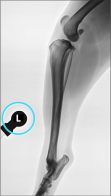
- Take the X-ray projection with the extended stifle
- Use a radiographic reference (blue circle) to adjust for radiographic magnification
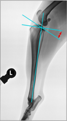
- Measure the TPA (= Tibial Plateau Angle)
- In patients with high TPA it is better to use locked plates.
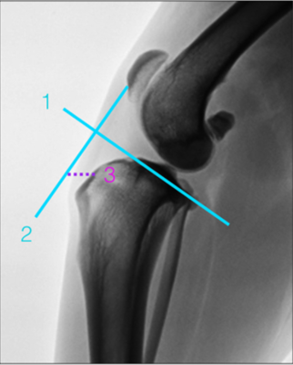
- Measuring the advancement of the crest
- Standard tibial plateau technique
- Draw the line for the tibial plateau (1)
- Draw the orthognal to (1) starting at the origin of the patellar tendon (PT) on the patella (2)
- Measure the distance (3) between the insertion of the PT on the tibial tuberosity (TT) and (2)
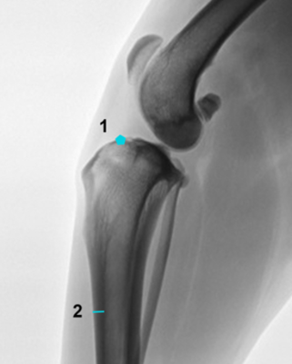
- Draw the position and direction of the osteotomy
- Mark a point immediately cranial to the tubercle of Gerdi (1)
- Measure the thickness of the tibial cranial cortex taking a point twice the length of the tibial crest starting from the TT (2)
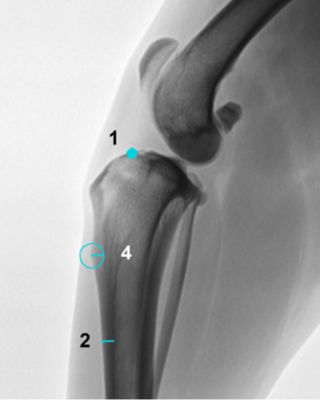
- Move the measurement (2) to the distal top of the tibial crest.
- Draw the circle (4) whose radius is the measure (2)
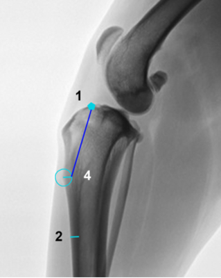
- Draw the line joining the point (1) with the midpoint of the arc of circumference (4) superimposed on the tibia

- Additional measures
- Measure the distance between the ALL and the cranial point (1) at the Gerdi tubercle
© X-Porous TTA created by Ad Maiora srl
Via della Costituzione 10, 42025, Cavriago – Italy
info@ad-maiora.eu
support@ad-maiora.eu
+39 348 868 3311
www.ad-maiora.eu
